When your immune system is fighting a cold, it makes itself known with stuffy noses, fevers, and coughs galore. Less conspicuously, it protects you when you bump your knee or scrape your arm (or worse). There is plenty of crosstalk between immunity and wound repair, indeed, and researchers are beginning to harness this relationship in various strategies for tissue regeneration. But in doing so, they are tasked with deconstructing the many convoluted mechanisms involved.
Neutrophils – “first responders” at the site of injury, if you will – are one of several immune factors that evoke positive and synergistic effects in wound repair. They work to stop pathogens in their tracks and recruit anti-inflammatory cells to the site. Monocytes and macrophages arrive soon after to clear out pathogens, dead cells, debris etc. while secreting molecules that move the healing process along.
Some immune factors are known to inhibit tissue regeneration, on the other hand. Toll-like receptors and interleukin-1 receptors are involved in signaling pathways that reduce the quality of wound healing, particularly in cases of ischemia-reperfusion and bone regeneration. Still, there is evidence that such receptors are involved in pathways that directly or indirectly support repair processes at the same time.
So we know that mechanisms of immunity can serve as positive or negative forces in tissue regeneration – or both. To complicate matters further, they are influenced by age and organ type among other factors. Julier et al suggest, nonetheless, that “the next generation of regenerative therapies may evolve from typical biomaterial-, stem cell-, or growth factor-centric approaches to an immune-centric approach.”
Regenerative medicine aims to restore function in cells, tissues, and organs by sometimes elaborate means. Below are two immuno-modulatory strategies covered in Julier’s review article:
Physiochemical Properties of Implants
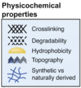
I am tempted to regard biomaterials as simply vehicles for therapy (e.g. cells, growth factors) and articles like this remind me otherwise. The implanted biomaterial contains powerful immune capabilities in and of itself. It consists of polymers with bonds, or cross-links, that link one polymer chain to another, and the degree at which this occurs impacts macrophage activity profoundly. For example, a higher cross-link density is known to elicit pro-inflammatory macrophages that secrete greater levels of cytokines. Surface chemistry certainly comes into play as well; hydrophobic, ionic surfaces are associated with more macrophages and less cytokines compared to hydrophilic, neutral counterparts. Figure 1 illustrates key physiochemical tools that can be modified at our disposal.
Extracellular Vesicles (EV)
EVs are little cargo bags released by cells for reasons that are as wide-ranging as they are innumerable. They are found in almost all cell types and all bodily fluids; traversing along the latter, they are “taken in” essentially by local or distant target cells. EVs from immune cells, in particular, are heavily speculated to be involved in the conversation between immunity and tissue healing by transporting critical immune factors to the site of injury. They can be loaded with exogenous agents and engineered to target specific cells for regeneration therapy. EV delivery methods are being explored, currently, ranging from intravenous injection to *ahem* biomaterial scaffolds.
Based on the aforementioned, incorporating the immune system into regenerative strategies is as exciting and inspiring as it is troubling. The concept isn’t entirely new, of course. The collection of evidence cited in this review article alone spans the better part of two decades. That being said, a suggested shift in therapeutic focus is avant-garde.
Google “regenerative medicine therapies” and you’ll find tons of info on growth factors, cells that secrete them, progenitor cells of cells that secrete them, and – you guessed it – biomaterials. Modulating the immune system gets a special shout out here and there, and that could very well amplify as evidence accrues. I guess only time will tell.