
The BMES blog posted recently about using multiphoton microscopy to study atherosclerosis, happening now at Cornell University. If you’re not familiar with atherosclerosis, you should be. It refers to the narrowing of arteries due to plaque buildup, and it will probably affect you or someone you love at some point.
This isn’t meant to frighten you; almost everyone over 60 has it to some extent, often without realizing. So when does atherosclerosis become critical? Heart disease is the short answer, my friends, the leading cause of death across developing and developed countries alike. Heart disease is pretty damn scary.
First, let’s lay down some facts from the publication:
- Who: Researchers at Cornell University led by Nozomi Nishimura
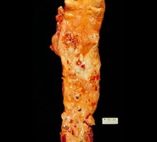
- What: Used three-photon microscopy (3PM) to image atherosclerotic plaque samples from humans and mice
- Why: To exploit third harmonic generation (THG), a signal that lets us see the cells and lipid deposits in lesions clearly. Current standard techniques do not accomplish this. THG is uniquely associated with 3PM, by the way.
- How: Diseased and normal tissue samples were obtained from murine aortas and human coronary arteries. Whole-mount (not sectioned) samples were imaged, sectioned for histology, and analyzed.
Now onto why this is unprecedented 🙏🏽
It harnesses revolutionary technology. First author David Small is a post-doctoral researcher in the Schaffer-Nishimura lab at Cornell University. “This technology can image deeper into tissues than previous multiphoton imaging lasers,” he explained to me in a pleasant exchange of emails. “There is often a trade-off between imaging depth and picture resolution, however this technology is bringing this trade-off closer together.” 3PM depicts cell and lipid morphology in great detail, including sub-cellular changes during early plaque formation.
This study is predicated on advances in technology led by Chris Xu – and prior to that – Winfried Denk, Watt Webb, and Jim Strickler. All of this happened at Cornell, of course, which I find incredibly endearing.
It’s simple. 3PM/THG uses a single laser that is commercially-available and user-friendly. Yes, other technologies can image atherosclerotic plaque to a likewise striking level of detail. Such systems often require the combination of different laser sources along with great expertise in setup and use, which is often impractical.
It can be used in vivo. Imaging with THG is label-free, which means the tissue is not injected with toxic contrast agents (aka no side effects). There is a pressing need for effective, less invasive technologies to monitor plaque formation in vivo, and 3PM could fill this void. “These early fat deposits can often correct themselves over time, however knowing why some do while others do not, would be something that this technology could investigate,” said Small.
The paper implies vast clinical potential for this tool. Along those lines, how would this be applied in the clinic? I posed the question to David Small: “Ideally this type of imaging would be attached to a intravascular endoscope.” While relatively invasive, according to Small, this is similar to currently-used techniques such as intravascular optical coherence tomography (OCT). “We would need to know that we can determine the extent of the pathology to a better degree than what is currently used.” With increased development, 3PM could very well be state of the art in understanding and diagnosing atherosclerosis.
I thanked David for what he does day in and day out, which had me quite emotional if I’m being honest. I recently lost someone I love very much because, well, heart disease. On that note, observing painstaking research that strives to reduce its morbidity and mortality has been uplifting. Seriously.
In a final comment, David reflects: “We think that this will lead to a greater uptake by biomedical researchers who don’t necessarily possess specific laser or imaging expertise, which in turn will advance the field of atherosclerosis research – and we both know how important that is.”

This is a very interesting application. Thanks for sharing!
LikeLike